- Time
- Topic
- Speaker
- Moderator
- 16:00 - 16:30
-
Cellular therapies in Parkinson's disease: recent advances and future directions
- Speaker:
Eng-King Tan
- Moderator:
Ruey-Meei Wu
- Time
- Topic
- Speaker
- Moderator
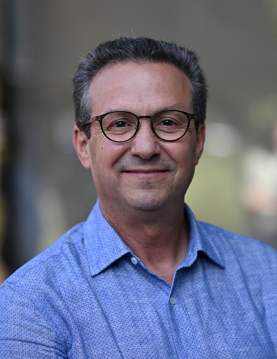
- Abdelhamid Benazzouz
- PhD
-
Research Director, Team Leader, Neurodegenerative diseases Institute, Bordeaux University
President / founder , Deep Brain Stimulation Society
E-mail:abenaz@u-bordeaux.fr
Executive Summary:
Abdelhamid Benazzouz is a Neurophysiologist Researcher employed by the Inserm Institute working in Bordeaux University. He is expert in the field of Neuroscience and especially in Parkinson’s disease. During his PhD, he pioneered in the discovery of high frequency stimulation of the subthalamic nucleus in a non-human primate model of Parkinson’s disease. Then he moved to Grenoble to participate in the transfer of this neurosurgical therapy to patients. In parallel with his hospital activity as a Neurophysiologist performing the electrophysiological mapping during surgery, he is the head of a research team investigating the functional mechanisms of DBS and the involvement of the monoaminergic systems in the pathophysiology of the non-motor symptoms of Parkinson’s disease. Abdelhamid Benazzouz was elected to be member of the National Scientific committee of CNRS (2012-2021). He co-founded two scientific Societies: The “Mediterranean Neuroscience Society” in 2009, for which he was the treasurer since 2015, and the “Deep Brain Stimulation Society” in 2021 for which he is the president Elect.
- Time
- Topic
- Speaker
- Moderator
- 17:00 - 17:30
-
Exercise as Neuroprotection in Early‑Stage Parkinson’s Disease: From Evidence to Implementation
- Speaker:
Tran Ngoc Tai
- Moderator:
Juei-Jueng Lin
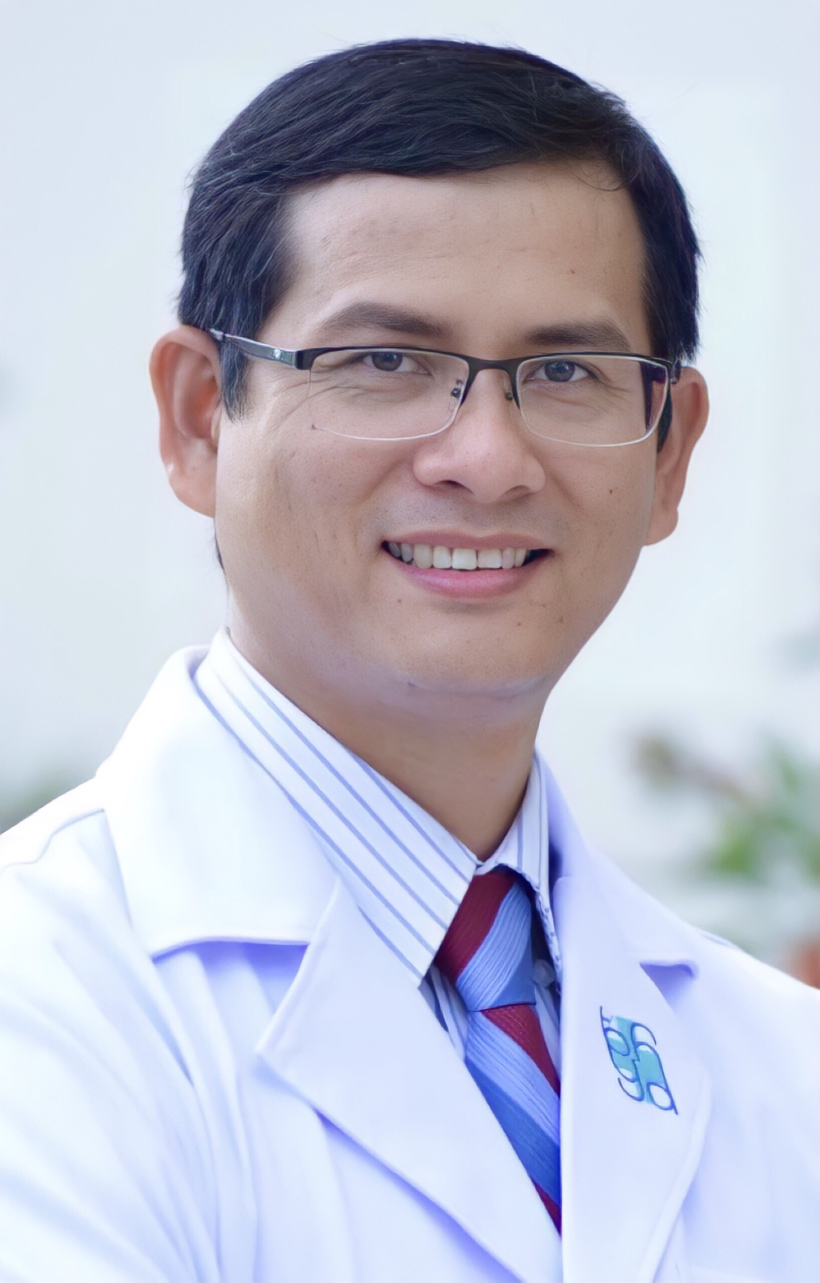
- Tran Ngoc Tai
- MD, PhD.
-
Head of Movement Disorder Unit, Deputy of Neurology Department, University Medical Center HCMC
President of Vietnamese Parkinson's disease and Movement Disorder Society, Vietnam Neurological Association
E-mail:tai.tn@umc.edu.vn
Executive Summary:
Tai Tran, MD, PhD, is a consultant neurologist and movement disorders specialist. He is the Head of the Movement Disorders Unit, Department of Neurology, University Medical Center, University of Medicine and Pharmacy at Ho Chi Minh City, Vietnam, and currently serves as the President of the Vietnamese Parkinson’s Disease and Movement Disorders Society under the Vietnam Neurological Association. His interest in movement disorders began in 2008. In 2013, he completed a six-month Deep Brain Stimulation training course at the Asia-Pacific Centre for Neuromodulation (ANCP), The University of Queensland, Brisbane, Australia, under the supervision of Professor Peter Silburn. He obtained his PhD degree in 2018 with a dissertation entitled “Botulinum toxin in the treatment of cervical dystonia”. His clinical and research interests focus on movement disorders, botulinum toxin therapy, and neuromodulation.